


 338 476

positive posterior margin
( Table 3). Cancer volumes at MRI
for the 13 patients with no recurrence and for the 4 patients
who recurred and whole-gland volume are shown in
Supplementary Figure 2.
Salvage robot-assisted RP resulted in undetectable PSA
<
0.1 ng/ml in three of four patients, durable over a
mean follow-up of 6 yr. Case 10 had a detectable PSA at
3 mo after completion of RP. There were no intraoperative
[(Fig._5)TD$FIG]
Fig. 5 – Case 10 (prostate-specific antigen [PSA] 7.24 ng/ml; prostate volume 27 cm
3
). Isolated anterior 2.6-cm
3
lesion suspicious at magnetic resonance
imaging in the anterior fibromuscular stroma on the midline and anterior right transition zone lobe (arrows): (a) T2; (b) apparent diffusion coefficient
map; (c) dynamic contrast-enhanced sequences. Targeted biopsies were positive for 8-mm Gleason score (GS) 7 (4 + 3) cancer. At histology, cancer was
at the anterior and inferior part of the specimen: 4.61-cm
3
volume, GS 7 (3 + 4), pT3a, pN0, and R1. Positive margins were anterior 6 mm and lateral
6 mm. Postoperative PSA was 0.41 ng/ml at 3 mo and 1.16 ng/ml at 18 mo postoperatively. Biopsies at left anterior part of preserved PZ showed
residual cancer on 3 mm GS 6 (3 + 3). Radical prostatectomy was performed at 2 yr with negative margins and detectable PSA.
[(Fig._4)TD$FIG]
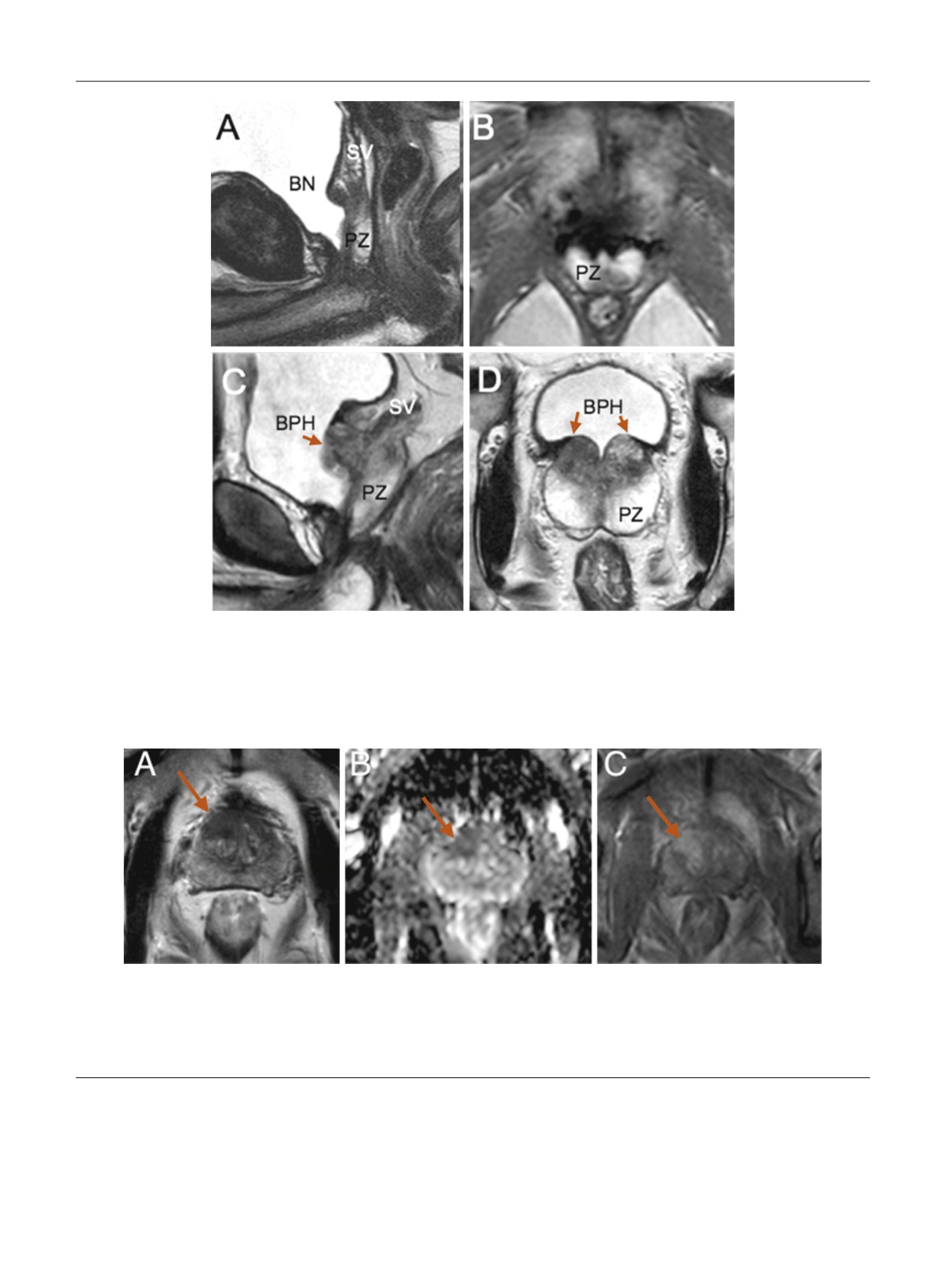
Fig. 4 – Cases 4 and 5 showing postoperative T2 magnetic resonance imaging (MRI) sequences at 6 mo, (a) sagittal and (b) transverse, and at 4 yr, (c)
sagittal and (d) transverse; preserved peripheral zone (arrows) and seminal vesicles. Case 14 (a, b) had stable prostate-specific antigen (PSA) of 0.74 ng/
ml at 6 mo and 0.70 ng/ml at 2 yr. Case 5 had rising PSA of 0.71 ng/ml at 6 mo and 1.34 ng/ml at 2 yr. MRI showed a symmetric area of benign
prostatic hyperplasia recurrence/persistence at the prostate base on each side of the bladder neck that may explain this PSA rise with time. Protocol-
based systematic biopsies were negative, and MRI was nonsuspicious for cancer.
BN = bladder neck; BPH = benign prostatic hyperplasia; PZ = peripheral zone; SV = seminal vesicle.
E U R O P E A N U R O L O G Y 7 2 ( 2 0 1 7 ) 3 3 3 – 3 4 2
338